

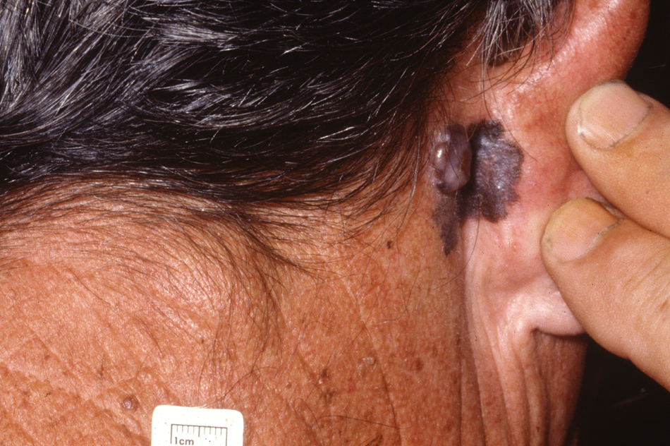
Head and Neck Melanoma
Dr John Chaplin. Head and Neck Specialist. Auckland
NECK AND NECK MELANOMA
Head and neck melanoma is complex and can present in a number of ways. Firstly as primary lesions of the skin of the face, scalp and neck and also as mucosal lesions of the mouth and sinonasal tract. It ca also present as metastatic disease to the lymph nodes in the parotid gland and neck.
The different types of malignant melanoma include:
- Superficial spreading melanoma most common, begins with initial radial growth phase prior to invasion
- Lentigo malignant melanoma : long radial growth phase, lentigo maligna is the precursor lesion.
- Acral lentiginous: most common form in darkly pigmented patients, often on the palms, soles, mucosal surfaces, nail beds (subungual melanoma). Often more aggrssive biological behavious.
- Nodular Melanoma: poor prognosis due to invasive nature from the onset.
- Mucosal Melanoma: Poor prognosis with late presentation and invasive potential
It is well documented that the lifetime risk of developing malignant melanoma has been steadily rising over the past several decades. For example in the United States, by the year 2000, approximately 1 in every 75 Americans was affected by this condition compared to 1/1500 in the early 1930s. However, interestingly enough, with extensive public awareness of the severity of melanoma, the mortality rate from melanoma even though gradually increasing, does not correlate with the rise in incidence. Early detection and diagnosis seems to profoundly effect the prognosis.
Diagnosis
Good history taking as well as careful and thorough examination of a suspicious skin lesion is important, but a definitive diagnosis of malignant melanoma depends on adequate biopsy sample. The method of choice in the current literature is excisional biopsy. A total excisional biopsy allows both the surgeon and pathologist to assess thickness, adequacy of excision margin and histiological characteristics of the lesion. These features are vital to determine whether or not further definitive surgery is needed to minimize risk of local recurrence.
However, if excisional biopsy is not possible ( lesion too large, unsuitable anatomical site ), the options include incisional biopsy, punch biopsy or saucerization.
Staging
Clarks and Breslow conducted studies that clarified the fact that the potential for metastatic spread is related to the depth of the melanocytic lesion from its epidermal origin. Table 1 looks at Clarks levels of invasion of malignant melanoma. Breslow measured tumour thickness from the top of the granular zone of the epidermis to the neoplasm base.
Level/ Depth of Penetration
I in situ ( confined to epidermis)
II extends into papillary dermis
III extends into juntion of papillary / reticular dermis
IV extends into reticular dermis
V invasion of subcutaneous tissue
Thickness is of main consideration in melanoma staging. The current system is largely based on the revised system of melanoma staging by American Joint Committee on Cancer (AJCC).
Revised Staging System for Malignant Melanoma [AJCC] Stage Criteria TNM
IA localized melanoma <0.75mm or level II T1N0M0
IB localized melanoma 0.76-1.5mm or level III T2N0M0
IIA localized melanoma 1.5-4mm or level IV T3N0M0
IIB localized melanoma >4mm or level V T4N0M0
III nodal metastases of only one regional lymph node basin any T,N1M0
or fewer than 5 in-transit metastases in absence of nodal
disease
IV advanced regional or distant metastases any T, any N, M1 / 2
Primary Treatment
The key factor in treating malignant melanoma is adequate excision of the primary lesion. It is well known that inadequate removal of a suspiciously melanocytic lesion leads to a high rate of recurrence. Excison of normal skin beyond the perimeter of the main lesion is recommended . There is ongoing debate of how deep and wide‚ the excision should be. Randomized studies conducted in the recent years suggest :
- Lesions classified as melanomas in situ is appropriately managed just by simple excision.
- Melanomas < 1.0mm thickness needs 1.0cm excision margins
- Intermediate lesion [1.0mm-4.0mm] needs 2.0cm excision margins
- Lesion greater than 4.0mm needs margins greater than 2.0cm.
The main consideration in performing a definitive excision is to minimize local recurrence. However, site is important in this consideration and a surgeon must consider the functional and cosmetic aspects particularly in the head and neck. Margins need to be adjusted accordingly.
Melanoma type is also imporatnt ie. It is widely accepted that malignant melanoma in situ with appropriate management has almost 100% 5 year survival rate. a wide excision is not justified in this instance.
The presence of satelitosis / satelite metastases is a poor prognostic sign. They reflect the ability of the malignant cells to survive at a distant site. The presence of microscopic satellites significantly decreases patients survival.
Head and Neck Melanoma Node Positive Disease
A patient presenting with metastatic disease to regional lymph nodes has a 50% chance of developing systemic metastases. Management of patients with postive nodal disease involves removal of the lymph nodes by neck dissection often combined with a parotidectomy if the parotid lymph node bed is either involved or at risk of having occult nodes from the site of the index primary lesion. Post surgical radiotherapy is usually recommended because of the high incidence of extranodal extension of tumour.
Elective Lymph Node Dissection (ELND)
In majority of patients, the regional nodes are clinically negative at the time of initial diagnosis, but may harbor occult micrometastases. ELND was developed as a therapeutic modality to prevent subsequent regional and distant spread should occult nodal involvement was present in the first place. The rationale of this treatment is based on the hypotheses that malignant melanoma will first metastasise to regional lymph nodes and only then to distant sites.
A large study involving 4682 patients by Slingluff et al found significant incidence of metastases to contralateral nodes (10%) and atypical nodal basins (6%) among those with nodal metastases. 3550 of patients were nodes-negative clinically at the initial point of diagnosis but 911 underwent ELND. This study found that only 16% overall of the ELND were positive. ELND did not prevent recurrent nodal disease in the dissected basin. This came from the fact that of the 911 subjects who underwent ELND, only 214 had nodal metastases (143 at the time of ELND, 71 at a later date). From the 71 subjects, 44% (31 patients) had nodal metastases in previously dissected basin. This results made the therapeutic value of ELND questionable. It is nonetheless important to remember that this study did not consider the thickness of the primary tumour. For example, they did not consider the thickness of the initial tumour in the 31 patients who had recurrent nodal metastases post-ELND.
Detractors of ELND argue that thin melanomas (<0.76mm) have virtually no risk of node micro-metastases and thick melanomas (>4.0mm) while having high risk of node metastasis, also have high risk of systemic spread. Neither would then benefit from ELND with respect to overall survival.
Balch et al in another study aimed to decide whether ELND would improve survival rate for patients with intermediate melanoma and also which subgroups of melanoma patients would have higher survival rate with ELND. This was designed as a prospective randomized surgical trial involving 786 patients with AJCC stages 1 and 2 melanomas. They found that ELND improves significantly the survival rates in patients 60 years or younger with intermediate-thickness melanoma. ELND patients older than 60 years old actually have a lower survival rate than those who only had node observation. The researchers where unable to ascertain if this was related to the immunosupressive effects of surgery or to other influences.
This study also supports the hypotheses of sequential metastasis of melanoma from the primary site to the regional nodes prior to other distant areas. It also suggests that there is a narrow window of time during which nodal micrometastases are isolated sufficiently that their removal prevents further dissemination.
The value of ELND remains controversial at this time. In summary, the current agreement is that · ELND is not justified in primary malignant melanomas in situ · ELND is not recommended for lesions in sites of ambiguous drainage such as the midline of head and neck · ELND should not be performed in elderly patients with co-morbid conditions, unless if the lesion lies directly over the nodal drainage · ELND is not justified in systemic metastases. In head and neck melanoma ELND would generally comprise a selective neck dissection of the at risk levels. See Neck Dissection
Sentinel Lymph Node Biopsy
A study by Reintgen et al in 1994 aimed at determining the order of melanoma nodal metastases. The investigators believed that melanoma is different from other solid tumours in terms of metastatic pattern. Most solid tumours (eg. breast cancer) show random nodal metastases pattern but melanomas is different because cutaneous lymphatic flow is better defined and can be mapped accurately by means of lymphoscintigraphy. This technique involves injecting a mean dose of human serum albumin (0.94 mCi) or technitium-99 intradermally around the the lesion, imaging using gamma camera set and obtaining dynamic flow study to identify the sentinel nodes. The sentinel lymph node is defined as the first node in the basin from which the primary site drained. The sentinel node is then harvested and assessed separately, this is then followed by complete node dissection. By comparing the nodal metastases between the sentinel and the nonsentinel nodes, the investigators came to a conclusion that there were no skip‚ metastases documented in any of the subjects. This particular study is not conclusive nor completely convincing because of the small number of patients. Further evidence from The MSLT1 trial has shown significant survival benefit (>26%) from SNB for intermediate thickness melanoma. We recommend SNB for all melanomas >1mm in depth particularly in head and neck melanoma.
Mucosal Head and Neck Melanoma
A rare from of head and neck melanoma that occurs primarliy in the nasal cavity and sinuses or, more rarely, in the oral cavity. Margins are limited by the surrounding structures and by the functional and cosmetic impact that wide surgical resections have. Regional lymph nodes are rarely involved in nasal cavity lesions but ELND is recommended in oral cavity lesions as occult metaststic rates are in the order of 30%.